Recently, the group of Image Guided Surgery and Therapy (IGST, Prof. Xin GAO) in Suzhou Institute of Biomedical Engineering and Technology (SIBET) proposed a publicly available standard image dataset and evaluation framework for assessing non-rigid 2D/3D registration algorithms (https://doi.org/10.5281/zenodo.997887).
In image-guided surgery, three-dimensional (3D) preoperative imaging with computed tomography (CT) or magnetic resonance imaging (MRI) is routinely used for surgery planning. However, the real-time 3D imaging information can hardly be obtained by CT or MRI during the surgery.
The intraoperative imaging equipment such as the fluoroscopy and portal imaging devices (PID) can only obtain the real-time 2D image. By registering the intraoperative 2D image and preoperative 3D image to recover the lost 3D spatial information in real-time, the intraoperative lesion dynamic tracking or positioning can be achieved.
For the present, the publicly available standard image datasets for 2D/3D registration are built based on the images of spine, lumbar, pelvis of human cadaver or the head of pig cadaver, which are not suitable for the non-rigid objects, such as lung.
In this research, a plasticized, inflatable pig lung model was used to obtain the image dataset. Inflated with different amounts of oxygen to simulate different respiratory phases, a sequence of 3D volume data was acquired by CT. With the lung model inflated and kept in certain states, 3D CT, 2D CT scout image and 2D X-ray were acquired for the same respiratory phases.
A total of 120 well-distributed landmarks in every 3D volume were manually annotated and checked by several radiologists.
Finally, an existing non-rigid 2D/3D registration algorithm was successfully evaluated based on the proposed image dataset and evaluation framework.
This research proposed a publicly available standard image dataset and evaluation framework for assessing non-rigid 2D/3D registration algorithms for the first time, which could facilitate the development and the clinical application of this advanced technique.
These works entitled “Thorax x‐ray and CT interventional dataset for nonrigid 2D/3D image registration evaluation” were published in Medical Physics (2018).
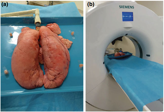
Fig 1. The image acquisition equipment. The pig lungs in the BioQuest Inflatable Lung kit (a) were fixed on a scanning table and connected to an oxygen tank during image acquisition (b).
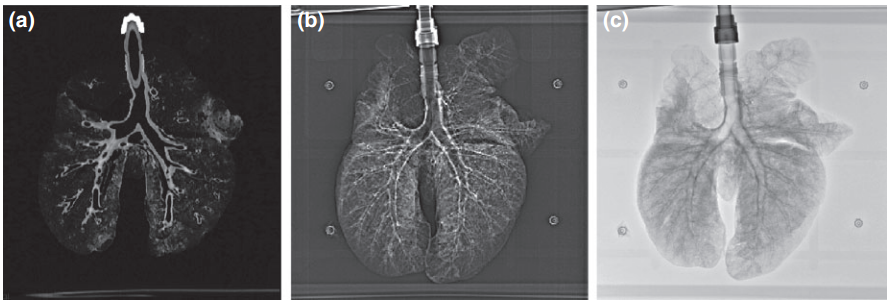
Fig 2. Images from the dataset.(a) CT image;(b) CT scout image;(c) 2D x-ray image.
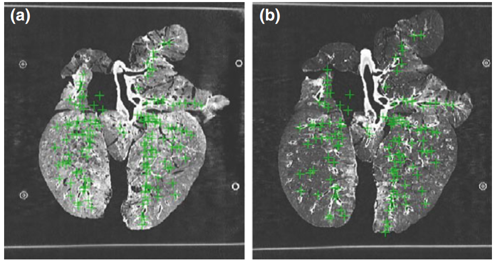
Fig 3. The annotated landmarks. Figure (a) and (b) show the landmarks (green crosses) in the end expiration phase and the end inspiration phase, respectively. Since all landmarks are projected onto a single slice, some of the green crosses are displayed outside the lung area. (Image by XIA Wei)
Keywords: 2D/3D registration; evaluation; image‐guided surgery; standard dataset.
Contact
XIAO Xintong
Suzhou Institute of Biomedical Engineering and Technology, Chinese Academy of Sciences (http://www.sibet.cas.cn/)
Phone: 86-512-69588013
E-mail: xiaoxt@sibet.ac.cn