Recently, the group of Image Guided Surgery and Therapy (IGST, Prof. Xin GAO) in Suzhou Institute of Biomedical Engineering and Technology (SIBET) proposed a preoperative radiomic model for noninvasively evaluation of tumor biological characteristics.(https://doi.org/10.1007/s00330-018-5763-x).
In recent years, the incidence of cancer in China has increased significantly. Cancer prevention and treatment has great social and scientific value.
Accurate analysis of tumor immunohistochemistry, gene expression, lymph node metastasis, differentiation levels and other biological characteristics contribute to the choice of the best treatment options, which can directly affect the prognosis outcomes.
Molecular markers for clinical expression of tumors need to be invasively sampled by surgery or needle biopsy, which can only be collected in a single position. This cannot fully describe the temporal and spatial heterogeneity of tumor tissues.
Imaging is commonly used as a clinical diagnostic tool that can be non-invasive, real-time, repeatedly obtain tumor information. Radiomics has been proposed as the integration of pattern recognition, machine learning and medical imaging diagnosis, which can be used to extract massive image features to solve the difficult problem in tumor assessment.
Tumor formation, metastasis and therapeutic response are affected by a variety of tumor biological characteristics. Therefore, it is of great significance to carry out multiple tumor biometric evaluations simultaneously in the same population.
In this study, magnetic resonance imaging data and tumor biological characteristics of 345 patients with rectal cancer were acquired (including immunohistochemical parameters such as Ki-67 and HER-2, lymph node metastasis, KRAS-2 gene mutation, and tumor differentiation).
Two feature selection methods and three classification methods were used. A non-invasive evaluation model for rectal cancer tumors was constructed.
The results show that the model has a good evaluation effect on tumor biological characteristics (AUC=0.651-0.720, Fig.1). The model can assess the tumor biological characteristics based on the MRI images (Fig.2), and may have good prospect in clinical application.
These works entitled "Preoperative Radiomic Signature based on Multiparametric Magnetic Resonance Imaging for Noninvasively Evaluation of Biological Characteristics in Rectal Cancer" were published in European Radiology.
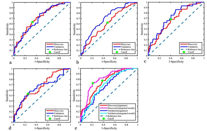
Fig 1. The receiver operating characteristic curves of radiomic signatures: (a) HER-2, (b) Ki-67, (c) tumor differentiation, (d) KRAS-2, and (e) lymph node metastasis. For lymph node metastasis, the ROC curves of integrated evaluation model were also plotted. (Image by XIA Wei)
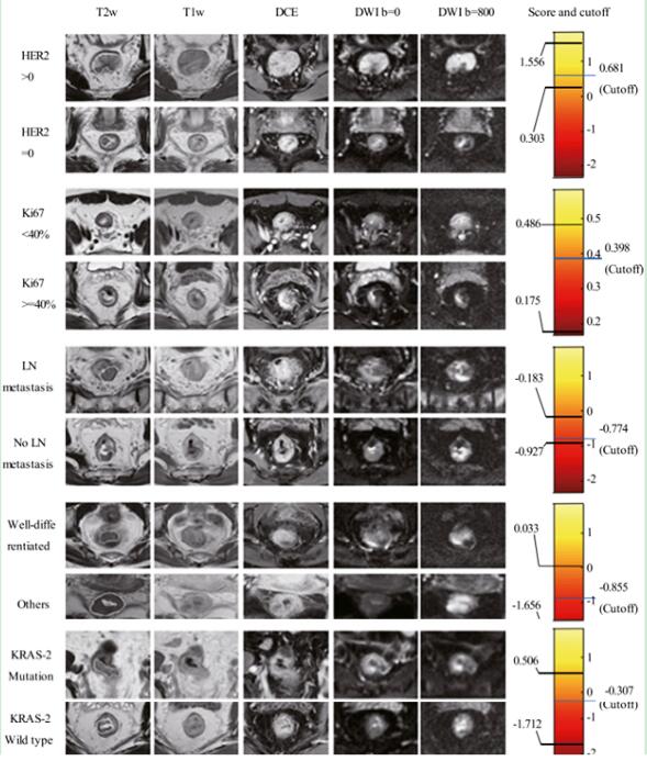
Fig 2. Preoperative images obtained by multiparametric magnetic resonance imaging and radiomic signature scores of patients with rectal cancer with different biological characteristics. (Image by XIA Wei)
Keywords: Rectal neoplasms, Magnetic resonance imaging, Radiomics
Contact
XIAO Xintong
Suzhou Institute of Biomedical Engineering and Technology, Chinese Academy of Sciences (http://www.sibet.cas.cn/)
Phone: 86-512-69588013
E-mail: xiaoxt@sibet.ac.cn