Brain’s functional repertoire and cognitive abilities are embedded in the details of the large-scale neural circuits that connect an immense number of neurons. Understanding how the nervous system collects, processes and stores information is not possible until we gain detailed descriptions of these circuits.
However, to date, a method that can depict complete circuitry of all neurons in a single brain tissue at neurite level is still in absence.
Researchers from the Suzhou Institute of Biomedical Engineering and Technology (SIBET) developed an optical multi-layer interference tomography (OMLIT) method that can image every cell in brain tissue with a large field of view at neurite level resolution.
The new method, developed by Dr. ZHANG Ruobing’s team at SIBET, provides high contrast images while keeps simplicity in hardware and operation.
The all-cell capturing capability of OMLIT lies in its multi-layer configuration of different materials (Fig.1).
ZHANG and his team collected serial ultrathin sections of resin-embedded brain samples at certain thicknesses of tapes that were selectively coated with a thin layer of reflective and conductive material.
The tape with sample sections was then laid down onto adhesive black carbon conductive tapes covering a silicon wafer. These layers of different materials form a multi-interface system where the interference among incident light, reflected field and scattering field generates high contrast images of all neurons and glial cells in the brain tissue.
Combinations of various materials and thicknesses of layers were carefully tested to achieve optimal imaging configuration, according to the researchers.
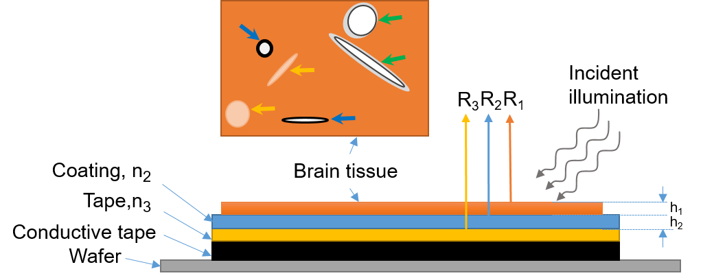
Fig.1 Schematic diagram of the multi-layer reflection model and cellular structures in a brain tissue sample. In the brain tissue layer, the blue arrows indicate the schematic drawings of axons, the green arrows mark that of blood vessels, and the yellow arrows indicate that of cell bodies. (Image by SIBET)
OMLIT is proved to be fast and accurate. It obtained a three-dimensional image dataset of mouse cortex (0.95×1.15×0.027mm3) at 360 nm pixel resolution within 12 minutes. One previous technique will need 3.5 hours to acquire the same volume with identical pixel size at 200 ns dwell time that produces similar image contrast.
The morphology and spatial locations of all neurons and glial cells, as well as the interweaving network of capillaries and backbone neurites can be distinguished and reconstructed from the OMLIT data (Fig. 2a).
Reconstruction accuracy was verified with serial SEM imaging of the same sample (Fig. 2b), as the two microscopies are seamlessly compatible.
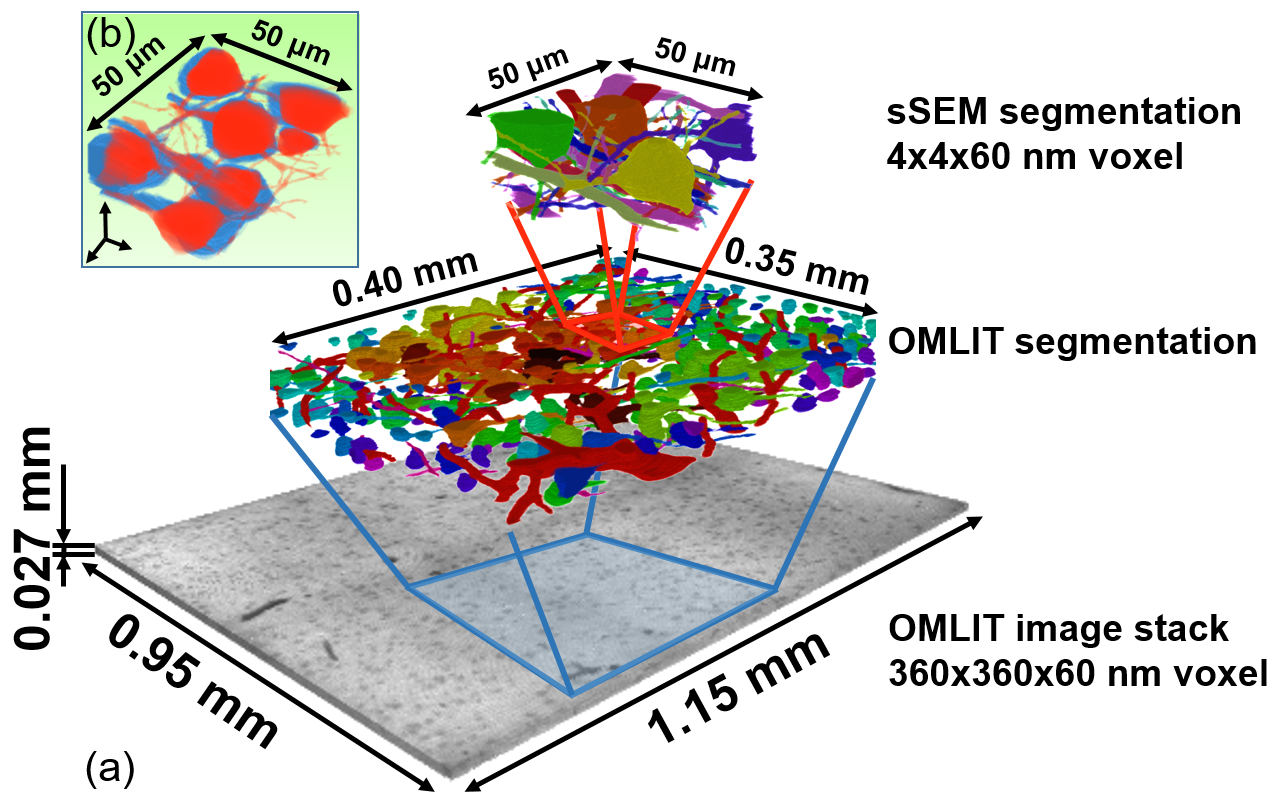
Fig.2 3D reconstruction of volumes of mouse cortex tissue by OMLIT and ATUM-sSEM. (Image by SIBET)
The technique, combined with ATUM- based serial SEM, “provides a promising approach to merge the complete map of long-range neural projections with complementary synaptic-level details of local microcircuits of all cells in a single brain tissue,” said ZHANG.
Further efforts such as automation of imaging pipeline and structure reconstruction are needed to foster higher-throughput implementation of OMLIT on larger-scale brain mapping.
The paper entitled “Optical Multilayer Interference Tomography Compatible with Tape-Based Serial SEM for Mesoscale Neuroanatomy” has been published online in ACS Photonics and was selected for supplementary cover.
Contact
XIAO Xintong
Suzhou Institute of Biomedical Engineering and Technology, Chinese Academy of Sciences (http://www.sibet.cas.cn/)
Phone: 86-512-69588013
E-mail: xiaoxt@sibet.ac.cn