Researchers at the Suzhou Institute of Biomedical Engineering and Technology (SIBET) of the Chinese Academy of Sciences recently proposed a hand-in-hand structured DNA assembly strategy, and developed an electrochemical/fluorescent dual-mode biosensor for circulating tumor DNA based on methylene blue and red-emissive carbon nanodots.
The new sensor, combines the characteristics of electrochemical and fluorescent sensors, whose signal sources and construction methods are usually quite different, according to the researchers.
Electrochemical sensor is a qualitative or quantitative method based on the correlation between target caused change of electrical signal and the concentration or other physical parameters. Fluorescent sensor is a qualitative or quantitative detection method by transmitting the information of the specific combination of target and the recognition element to the fluorescent element, causing the change of fluorescence intensity or emission wavelength.
"Integrating the two technologies for synchronous detection in one unique system can not only effectively improve the detection accuracy, but also reduce the influence of background signals, instrument fluctuations and other factors on the obtained signals," said MIAO Peng, leading researcher of the study and also a scientist from SIBEBT.
The sensor has a wide application prospect in basic research, environmental detection, clinical testing and other fields.
Probe A is immobilized at the electrode surface via the thiol group labeled at its 5’ terminal. In the presence of target, complete double strands are formed between target and its binding domain in Probe A.
Meanwhile, the stem region is opened and single-stranded region is released which is responsible to open the second hairpin of Probe B. It is previously conjugated with red emissive carbon nanodot through the 3’ terminal NH2.
Subsequently, the hairpin structure of Probe C is opened with the released single-stranded region of Probe B.
In addition, Probe C is able to displace target sequence to form a complete three-way junction structure. The target is thus recycled and assist the formation of multiple three-way junctions. Since abundant methylene blue molecules at the 3’ terminal of Probe C are located near the electrode interface, significant electrochemical response could be recorded to reveal target.
The released single-stranded swing arm formed by 3’ terminal of Probe A and 5’ terminal of Probe B on the three-way junction act as DNAzyme coupling 3’ terminal of Probe B on adjacent three-way junction as the substrate. Hand-in-hand structured DNA monolayer is thus fabricated.
With the existence of Mg2+, the substrate sequence can be cleaved and the conjugated carbon nanodots are released into the solution. "By measuring the increased fluorescence emission, original target level can also be evaluated by fluorescence technique," said MIAO.
Theoretical calculation and gel electrophoresis imaging confirmed the feasibility of the reaction(Figure 1). The synthesized carbon nanodots have strong anti-interference and can maintain high fluorescence stability in the physiological environment (Figure 2).
Through a series of condition optimization and quantitative tests, MIAO and his team established the linear calibration curves of electrochemical/fluorescence intensities and target concentration, which can achieve a wide linear range of six orders of magnitude.
At the same time, the content of target can also be easily distinguished by fluorescence imaging (Figure 3). The dual-mode sensor developed in this work is novel with high sensitivity and strong scalability, which can provide a powerful tool for nucleic acid analysis and clinical diagnosis.
Results of the study entitled “Hand-in-hand structured DNA monolayer for dual-mode analysis of circulating tumor DNA” were published in the latest issue of Chemical Engineering Journal.
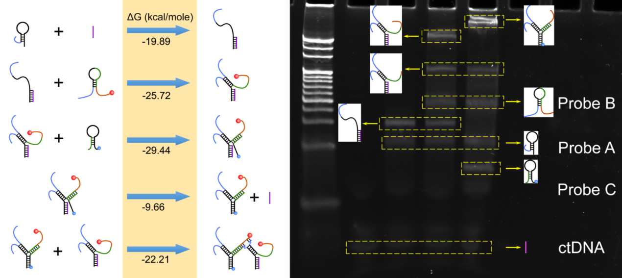
Figure 1. Standard free energy (ΔG) of reaction processes and PAGE analysis. (Image by SIBET)

Figure 2. Fluorescence responses of red-emissive carbon nanodots towards potential interfering ions. (Image by SIBET)
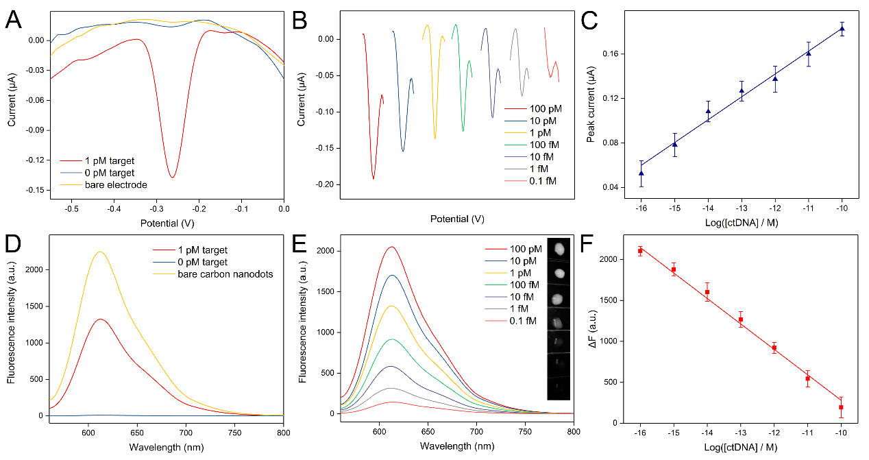
Figure 3. Square wave voltammograms and fluorescent responses after target DNA triggered DNA assembly. (Image by SIBET)
Contact
XIAO Xintong
Suzhou Institute of Biomedical Engineering and Technology, Chinese Academy of Sciences (http://www.sibet.cas.cn/)
Phone: 86-512-69588013
E-mail: xiaoxt@sibet.ac.cn