Zebrafish has been widely used in developmental biology, immunology, neurobiology, and drug screening and shows significant superiority in the large-scale heterogeneous studies of development and drug effects due to its small size, short life cycle, high fecundity, and low cost.
However, practical experiment processes are heavily dependent on hand manipulation and imaging under traditional microscopes, with large workloads but very low efficiency, which limits the utilization of Zebrafish in large-scale analysis.
Researchers at Suzhou Institute of Biomedical Engineering and Technology (SIBET) and Shanghai Institute of Nutrition and Health (SINH) of the Chinese Academy of Sciences (CAS) developed a light-sheet flow imaging system (LS-FIS) for high-throughput 3D imaging of zebrafish based on flow light sheet to address these bottlenecks.
Light-sheet microscopy (LSM) is a 3D imaging method with low phototoxicity and fast imaging speed. However, current light sheet imaging requires a complex sample preparation process for zebrafish imaging. Moreover, due to the limited field-of-view, 3D imaging of whole embryos often requires multi-region imaging and stitching, which severely limits the imaging flux of this technique.
By a smart design of fluid flow and optical coupling system, and using precise sampling control timing and 3D reconstruction algorithms, LS-FIS combines flow imaging technology with light sheet illumination to achieve high throughput 3D imaging of 200 embryos/hour.
LS-FIS was applied to study the development of zebrafish trunk and head vascular. 100 transgenic zebrafish Tg (kdrl: EGFP), in which their vascular endothelial cells labeled with green fluorescent proteins, were imaged by LS-FIS every day from 3 to 9 dpf for demonstration.
Over 500 3D images of whole embryonic zebrafish were obtained, and typical imaging results are shown below in figure 1. This is the first known report of large-scale whole-fish 3D imaging data.
The length of intersegmental vessels and changes in the morphology of the hyaloid vascular were measured and analyzed with the obtained 3D structures. Results show significant heterogeneity of the trunk vessel development but less heterogeneity of the hyaloid vascular.
The work entitled "Heterogeneities of zebrafish vasculature development studied by a high throughput light-sheet flow imaging system" was published in Biomedical Optics Express (V13(2022), pp. 5344-5357). The first author of this paper is Assistant Researcher YANG Guang, and the corresponding author is Professor LI Hui.
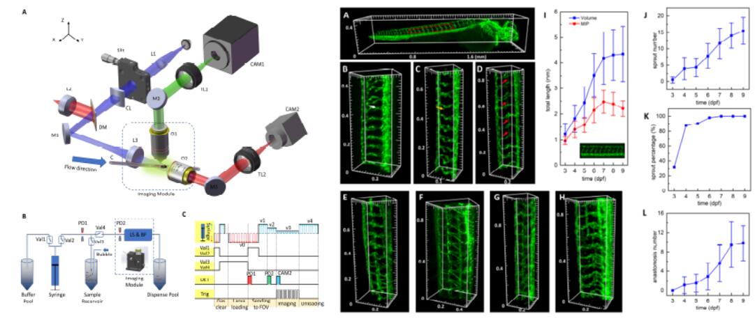
Fig. 1. Schematic diagram of the high-throughput 3D imaging system LS-FIS (left) and statistical study of the vascular development process from 3 to 9 dpf for hundreds of zebrafishes (right).
The prototype of LS-FIS has been validated for several iterations for better stability, appearance, and easy-to-use software interface. It was also tested in the SINH and Institute of Neuroscience of CAS and proved to have a good imaging capability and versatile usage.
The system was received extensive attention during its exhibition at the China Zebrafish Conference 2021 (Guiyang, Guizhou Province).
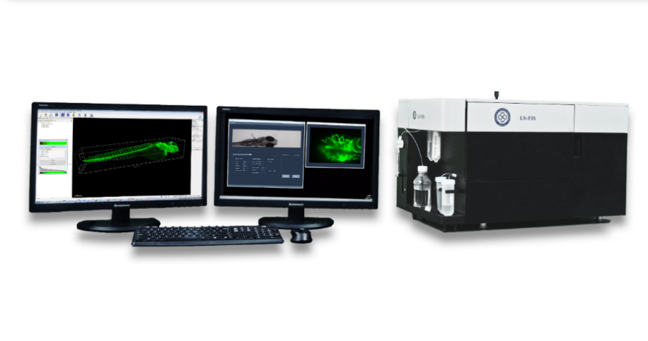
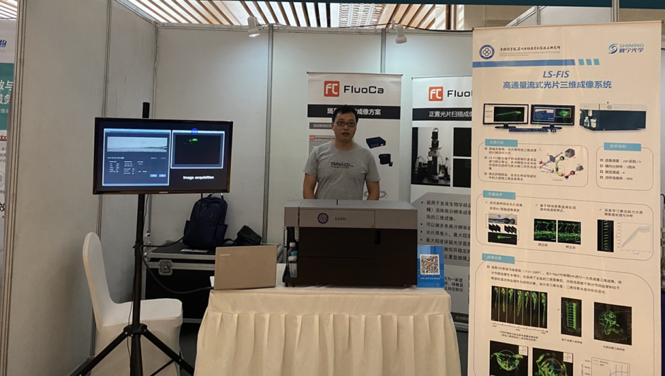
Fig. 2. Prototype of the high-throughput 3D imaging system LS-FIS (top) and exhibited at the China Zebrafish Conference 2021 (bottom).
The group further developed relevant image analysis and processing algorithms to deal with large amounts of 3D image data captured by LS-FIS. For zebrafish intersegmental blood vessels, they proposed a 3D convolutional neural network with multiscale features (MS-3D U-Net) to achieve segmentation and recognition of 3D vascular. By multiscale feature learning and an optimized loss function based on a hard attention mechanism, MS-3D U-net achieved an accuracy of over 90% (AUC value).
Based on MS-3D U-Net, the 3D image data of zebrafish embryos continuously observed for 24 hours were automatically segmented and measured, and the developmental curves of intersegmental vessels and dorsal longitudinal anastomosing vessels were plotted.
The work entitled "Optimized U-Net model for 3D light-sheet image segmentation of zebrafish trunk vessels" was also published in Biomedical Optics Express (V13(2022), pp. 2896-2908). The first author of the paper is graduate student YIN Jingyi, and the corresponding author is Assistant Researcher WANG Linbo.
This work was supported by the instrumentation and equipment development of Chinese Academy of Sciences, National Natural Science Foundation of China, and Suzhou National New & Hi-Tech Industrial Development Zone Leading Talents projects.
Contact
XIAO Xintong
Suzhou Institute of Biomedical Engineering and Technology, Chinese Academy of Sciences (http://www.sibet.cas.cn/)
Phone: 86-512-69588013
E-mail: xiaoxt@sibet.ac.cn