Photobiomodulation therapy, is one of the efficient and easily-accepted non-pharmacological treatment options for patients with knee osteoarthritis, a disease common among people over 60 years old.
However, most phototherapy devices currently ignore the analysis of the light dose delivered to the target tissue, which severely affects the therapeutic effect of photobiomodulation.
In a recent study published in the journal Biomedical Optics Express, researchers from Suzhou Institute of Biomedical Engineering and Technology (SIBET) of the Chinese Academy of Sciences developed a knee optical model. Based on this model, the researchers investigated the influence of light source parameters on the delivered dose to the deep joint during phototherapy for knee osteoarthritis. Based on this model, the researchers reproduced the phototherapy process for knee osteoarthritis and investigated the impact of treatment light source parameters on the dose reaching the deep joint tissues.
“Dosimetric analysis of knee osteoarthritis phototherapy and the establishment of a system for controlling and regulating the phototherapy dose for knee osteoarthritis are of great significance for the precise phototherapy of patients with knee osteoarthritis,” said XIONG Daxi from the Light&Health Center at SIBET, leading author of the study.
The researchers voxelized patients’ magnetic resonance imaging (MRI) data, classified and assigned values based on differences in optical properties, and then employed Monte Carlo simulation method to investigate the light transport in tissues.
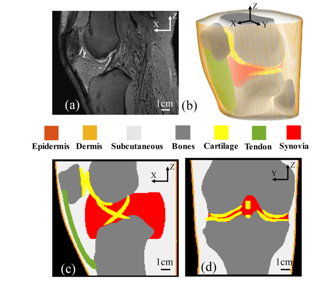
Fig. 1 (a) MRI image of knee joint; (b) Overall model image of knee joint; (c) Sagittal plane of the model; (d) Coronal plane of the model; In (b)-(d), to better distinguish the difference between the optical properties of the tissue, different colors are utilized to indicate tissues. (Image by SIBET)
Based on skin optical properties, XIONG and his team created a multi-layer skin phantom to characterize the epidermis, upper dermis, and deep dermis in the skin. They simulated the skin conditions of patients with different skin colors by adjusting the concentration of absorbent and scattering agent.
By comparing and analyzing the correlation of the incident light transmittance in the optical model and the created phantom, the team validated the reliability of the optical model they proposed.
“The optical properties of the skin in the knee joint model need to be finely adjusted for different patients to reduce the error introduced by differences in skin color among patients,” said XIONG.
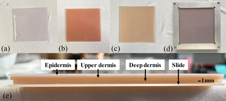
Fig. 2. (a)-(c) the phantoms of the epidermis, upper dermis, and deep dermis; (d) the top view of the three-layer phantom; (e) a side view of the three-layer phantom. (Image by SIBET)
They performed multiple optimization adjustments of the optical characteristic parameters in the optical model through knee joint correction experiments, so as to make the results of the optical model more closely aligned with the actual treatment situation of the patients.
Researchers selected different characteristic points on the knee as LED beam treatment areas and studied the effect of the light source irradiation position on the absorption of light dose by target tissues in the joint cavity.
Their result showed that the optimal irradiation position for deep treatment of knee osteoarthritis was on both sides of the patella, where there was the greatest light dose absorption. The optimal height of the light source was located at a medium-high position parallel to the knee joint space.
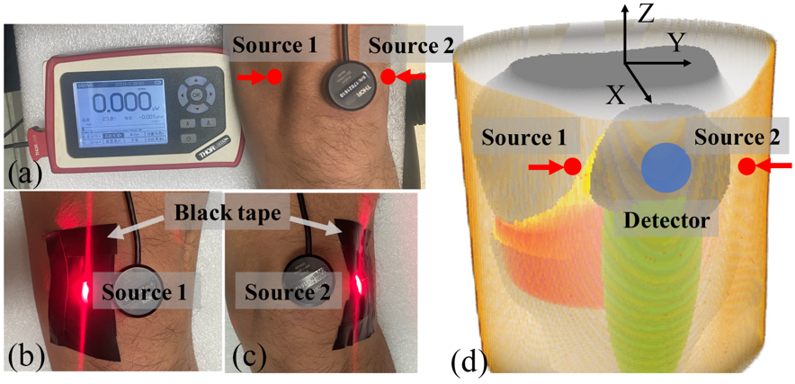
Fig. 3. (a) the photo of the left knee detection experiment using the optical power meter; (b) the irradiation at the position of Source 1; (c) the irradiation at the position of Source 2; (d) the irradiation position and detection position in simulation. Cover source with black tape can prevent source from spilling out in all directions and affecting the detection results. (Image by SIBET)
The method helps enable the study of subcutaneous light dose distribution, and further improve the model reliability through phantoms and correction experiments, according to XIONG.
The study offers optimization possibilities for existing knee osteoarthritis phototherapy products, and solves the problem of light dose measurement in the knee joint area, providing reference for the analysis of light therapy dose in other parts in the future, XIONG said.
Contact
XIAO Xintong
Suzhou Institute of Biomedical Engineering and Technology, Chinese Academy of Sciences (http://www.sibet.cas.cn/)
Phone: 86-512-69588013
E-mail: xiaoxt@sibet.ac.cn