Ovarian cancer has the second highest incidence and the highest mortality rates in gynecological malignancy in China. Epithelial ovarian cancer (EOC) accounts for 90 – 95% of all ovarian cancers. Standard treatment strategy for EOC consists of operative tumor debulking and administration of intravenous chemotherapy.
A team of researchers led by GAO Xin at Suzhou Institute of Biomedical Engineering and Technology adopted deep learning in EOC segmentation and evaluated its feasibility.
“Assessment of tumor prior to surgery is of paramount importance in guiding treatment decisions and ultimately improving patient outcomes,” said GAO. Identification and depiction of the tumor area (EOC segmentation) is a prerequisite for tumor evaluation, which also helps simplify subsequent efficacy assessment.
Nevertheless, EOC segmentation is commonly conducted in a slice-by-slice manner by clinicians in clinical practices, which is both time-consuming and labor-intensive. Hence, a solution to this problem is urgently required.
“With the rapid increase of medical image data, the need for fully automated EOC segmentation methods is becoming more and more urgent,” said HU Dingdu, first author of the study.
GAO and the team included a total of 339 magnetic resonance (MR) images of EOC patients from eight hospitals in their study. Five evaluation metrics were selected to assess the segmentation performance of different models.
Their results showed that U-Net++ achieved optimal segmentation results in both internal and external test sets demonstrating the superior accuracy and generalization of the model.
In addition, the team further analyzed the effects of tumor stage and histological type on the segmentation performance of the models.
However, the segmentation accuracy of the model on advanced stage tumors and serous tumors is relatively low, “indicating that different tumor stages and histological types have influence on the segmentation performance of the model”, according to GAO.
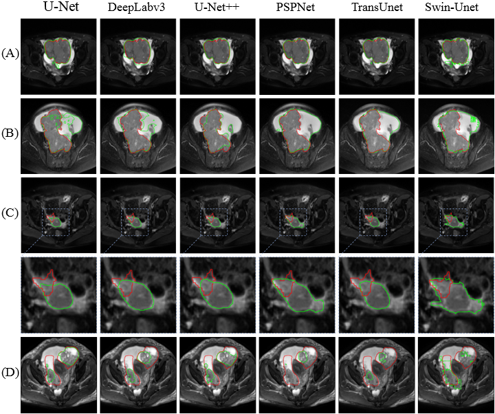
Figure 1. Visual comparison of segmentation results of different models. (Image by SIBET)
This study explored and validated the potential application of artificial intelligence techniques for fully automated segmentation of EOC. By integrating with the team's previous studies in differentiating epithelial ovarian tumors and type I and II EOC, an end-to-end fully automated EOC diagnostic process is highly potential for improvement of clinical efficiency.
The research was funded by institutions including the National Natural Science Foundation of China. The results were published in the journal Quantitative Imaging in Medicine and Surgery.
Contact
XIAO Xintong
Suzhou Institute of Biomedical Engineering and Technology, Chinese Academy of Sciences (http://www.sibet.cas.cn/)
Phone: 86-512-69588013
E-mail: xiaoxt@sibet.ac.cn