Researchers at Suzhou Institute of Biomedical Engineering and Technology (SIBET) of the Chinese Academy of Sciences recently designed a novel kind of red-emissive silicon nanodots (R-SiNDs) for mitochondrial dynamic tracking and blood-brain barrier penetration imaging.
As the main site of aerobic respiration in eukaryotic cells, mitochondria not only provide energy for the cells but also are closely related to some cellular activities. The lack of mitochondrial function can also lead in diabetes, cardiac arrhythmia, Alzheimer’s disease and many other diseases.
Fluorescent probes are regarded as the material of choice for mitochondrial imaging due to their good targeting and the advantage of real-time feedback. Nevertheless, organic dyes usually show weak resistance to photobleaching, and less favourable ability to penetrate the blood-brain barrier (BBB). DONG Wenfei and his team at SIBET adopted Silicon nanodots (SiNDs), an emerging nanomaterial with low/non-toxicity and environmentally friendly characteristics, to exploit new fluorescent probes with both BBB penetration ability and excellent optical properties for long-time imaging.
“Fluorescent materials with red emissions possess special advantages for imaging, such as lower phototoxicity, high tissue permeability, and lower interference of autofluorescence from organisms,” said DONG. “small particle-size SiNDs with positive surface charge and amphiphilic properties fulfill the basic requirements for penetrating the BBB”.
Their study showed that the R-SiNDs have excellent targeting and imaging ability towards living cell mitochondria. Moreover, R-SiNDs can be used as monitors to achieve the visualization of real-time mitochondrial dynamics due to their wonderful optical properties.
By the robust tools of R-SiNDs, the researchers also observed the fusion and division processes of mitochondria (Fig. 2a-c). Results showed that the lipid droplets (LDs) were free from above the mitochondria to below and subsequently wrapped by the mitochondria with the changing of mitochondria shape. The changes in the position of LDs and the morphology of mitochondria resulted in a larger contact area between the two organelles, which was more favorable for the process of material exchange.
More interestingly, R-SiNDs without any other modification exhibited wonderful blood-brain barrier (BBB) penetration capacity and were accumulated in the central canal of the spinal cord of zebrafish. “It is speculated that the small particle size, positive charge and amphiphilic properties together contribute to the BBB penetration by R-SiNDs, which also lays the foundation for the diagnosis of R-SiNDs in brain-related diseases,” said LIU Yulu, first author of the study.
The R-SiNDs can not only be used to study mitochondrial dynamics, but provide a reliable basis for diagnosing mitochondria-related brain diseases, which is of great value in biomedical applications, according to the researchers.
The research results entitled “Red-emissive silicon nanodots with highly biocompatible for mitochondrial dynamic tracking and blood-brain barrier penetration imaging” were published in Sensors and Actuators B: Chemical.
The research was supported by the National Key R&D Program of China, National Natural Science Foundation of China, etc.
Link to paper: https://doi.org/10.1016/j.snb.2024.135523
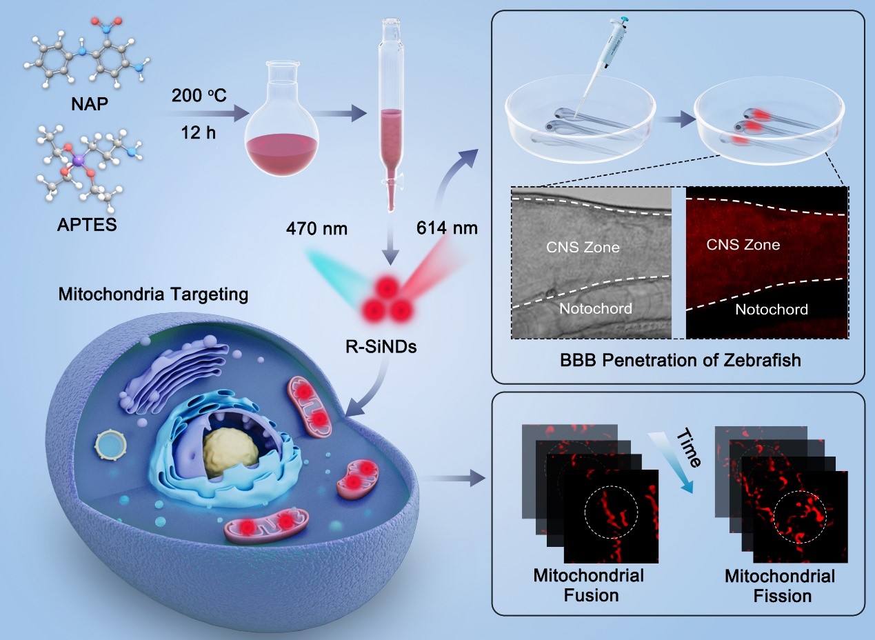
Fig.1. Schematic illustration for the synthesis of the R-SiNDs, and the application for long-time mitochondrial dynamics imaging and zebrafish neurological imaging. (Image by SIBET)
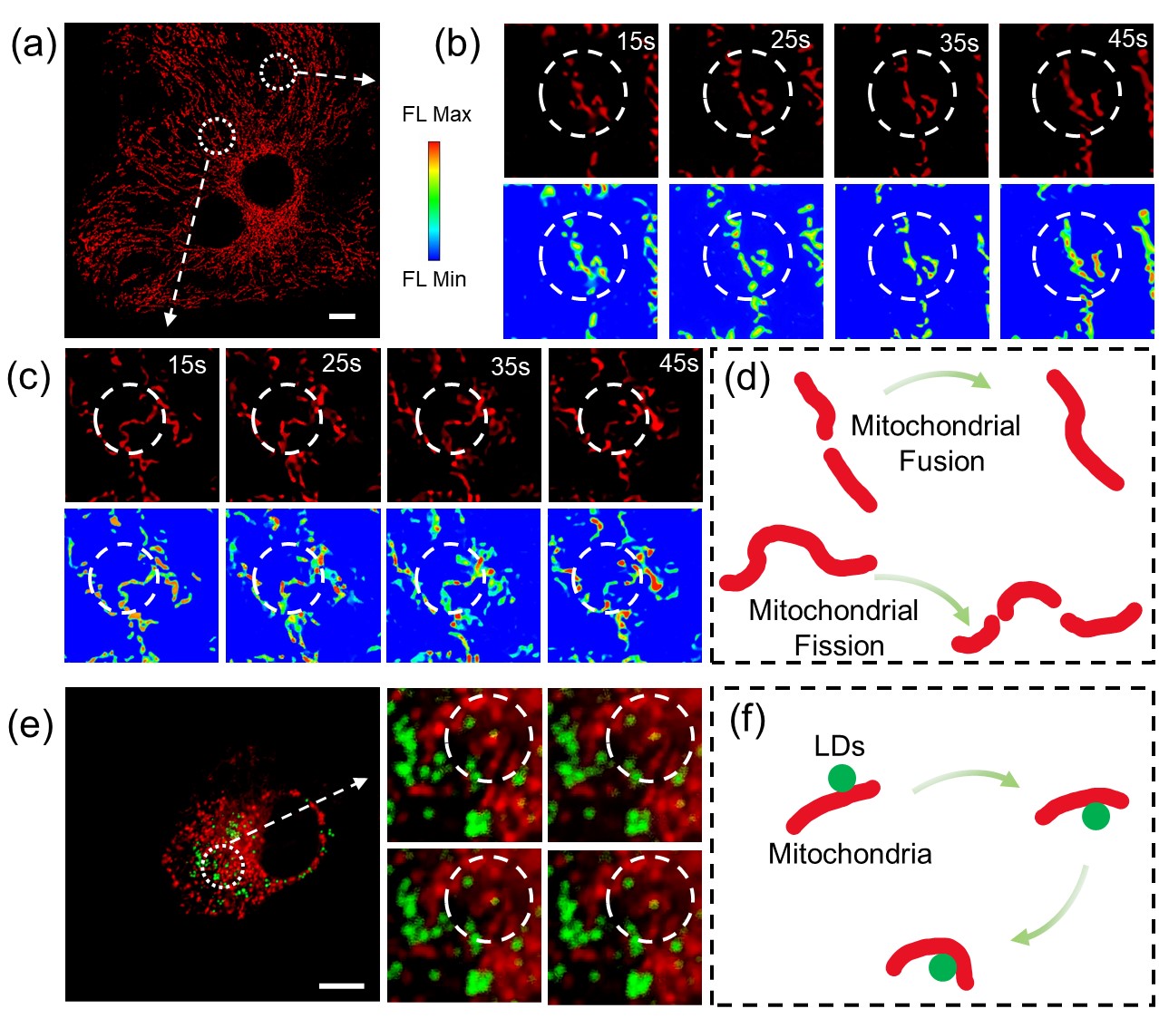
Fig.2. (a) Confocal microscopy images of mitochondria after staining PANC-1 cells with R-SiNDs. (b) The fusion process of mitochondria labeled with R-SiNDs. (c) The fission process of mitochondria labeled with R-SiNDs. (d) Schematic illustration of mitochondrial fusion and fission. (e) Dual-color confocal microscopy images of PANC-1 cells after co-staining with R-SiNDs and BODIPY. (f) Schematic illustration of the mitochondria parcels LDs. Scale bar: 10 μm. (Image by SIBET)
Contact
XIAO Xintong
Suzhou Institute of Biomedical Engineering and Technology, Chinese Academy of Sciences (http://www.sibet.cas.cn/)
Phone: 86-512-69588013
E-mail: xiaoxt@sibet.ac.cn